|
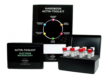
Actin-Toolkit Electron Microscopy, TEM용 actin 시료 준비
관련 단백질 연규에서 표적단백질이 actin에 미치는 영향을 확인할 때 전자현미경을 이용한 관찰은 매우
유용합니다. Actin-Toolkit Electron Microscopy는 TEM 분석을 위한 actin 시료를 준비하는 제품입니다.
시료는 negative staining하여 TEM으로 actin filaments의 사이즈 및 구조관찰을 할 수 있습니다.
The Actin-Toolkits Electron Microscopy is available with α-actin (rabbit skeletal muscle and
bovine α-cardiac actin.
The kit is used for electron microscopic analysis of negatively stained proteins and protein complexes.
The Toolkit is ready-to-use for the preparation of of electron micoscopic specimen,making this method
accessible for researchers who like to get introduced to EM works on G- and F-acton.
Applications
• Molecular imaging analysis of ligands bound to monomeric actin or actin filaments, bundles
or networks by TEM
• Determination of length, thickness or properties of actin filaments
사용 예
 |
|
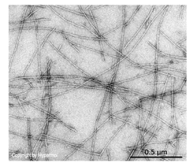
Electron Microscopy of actin filaments negatively
stained with 1% uranly-acetate.
|
 |
|
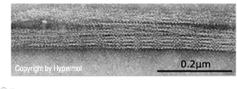
Electron Microscopy of actin filament bundles
(paracrystals) induced by addition off 50mM
MgCl2 to actin filaments. Negatively staining
with 1% uranly-acetate.
|
Rabbit Skeletal Muscle Alpha Actin
Description
|
Protein
|
|
Actin
|
|
Origin
|
|
Skeletal muscle alpha actin, rabbit
|
|
Molecular mass
|
|
42kDa
|
|
Toolkit description
|
|
Electron microscopic analysis of negatively stained proteins a highly evident
method to demonstrate the effects of ligands on actin. The Actin-Toolkit Electron
Microscopy is ready-to use for the preparation of electron microscopic specimen,
making this method accessible for researchers who would like to get introduced
to EM works on actin imaging.
This Toolkit is designed for users that either took a short introduction at an EM
facility or to pass kit and ligands for inspection to an EM service. The Actin-Toolkit
Electron Microscopy is easy-to-use. It contains 4x0.5mg rabbit skeletal
(Cat. #8070-01) or bovine cardiac muscle actin (Cat # 8700-01), buffers for washing
the specimen that maintain either the G- or F-actin state, and SpreadingSolution
in addition.
A handbook with a detailed description of the experimental seyup guides the user
for safe, error-free handling. We guarantee excellent performance of kit componenets
when stored at -70°C upon arrival for 6 months.
|
|
Purity
|
|
>99% by scanning densitometry
|
Properties
|
Form
|
|
Lyophilized, ready-to-use
|
|
Kit content
|
|
4x0.25mg G-Actin (lyophilized powder)
4x50ml MonoMix (lyophilized G-actin ATP-buffer)
2x1.0ml PolyMix 10x stock (lyophilized F-actin ATP-buffer, pH 7.4)
1x0.5ml SpreadingSolution
1x0.5ml 1M MgCl2
4x Centrifuge-Miniffilter (0.2µm)
1x Toolkit Handbook Electron Microscopy
|
|
Actin Buffer
|
|
2mM Tris-Cl pH 8.2, 0.4mM ATP, 0.5mM DTT, 0.1mM CaCl2, 1mM NaN3 and 0.3%
disaccharides, when reconstituted with 1.0 ml ultrapure water to obtain a 1mg/ml
solution.
|
|
Purity
|
|
>99% (according to scanning densitometry)
|
|
Purification notes
|
|
Purified from rabbit skeletal muscle, GPC.
|
|
DOL
|
|
Determined by Biuret method.
|
|
Storage instructions
|
|
We guarantee stable performance of the kit components for 6 months when stored at
-70°C upon arrival. Solubilized G-actin is kept on ice and should be used within 1
week. Avoid refreezing.
|
|
Shipping conditions
|
|
At ambient temperature. Upon delivery store at -70°C.
|
|
Remarks
|
|
For Use in Research only. Not for Use in Human or Veterinary Diagnostical or
Therapeutical Applications.
|
|
|
|
|
Alpha Cardiac Actin, Bovine
Description
|
Protein
|
|
Actin (alpha-cardiac actin)
|
|
Origin
|
|
Cardiac muscle, bovine
|
|
Molecular mass
|
|
42kDa
|
|
Toolkit description
|
|
Electron microscopic analysis of negatively stained proteins a highly evident
method to demonstrate the effects of ligands on actin. The Actin-Toolkit Electron
Microscopy is ready-to use for the preparation of electron microscopic specimen,
making this method accessible for researchers who would like to get introduced
to EM works on actin imaging.
This Toolkit is designed for users that either took a short introduction at an EM
facility or to pass kit and ligands for inspection to an EM service. The Actin-Toolkit
Electron Microscopy is easy-to-use. It contains 4x0.5mg rabbit skeletal
(Cat. #8070-01) or bovine cardiac muscle actin (Cat # 8700-01), buffers for washing
the specimen that maintain either the G- or F-actin state, and SpreadingSolution
in addition.
A handbook with a detailed description of the experimental seyup guides the user
for safe, error-free handling. We guarantee excellent performance of kit componenets
when stored at -70°C upon arrival for 6 months.
|
|
Purity
|
|
>99% by scanning densitometry
|
Properties
|
Form
|
|
Lyophilized, ready-to-use
|
|
Kit content
|
|
4x0.25mg G-Actin (lyophilized powder)
4x50ml MonoMix (lyophilized G-actin ATP-buffer)
2x1.0ml PolyMix 10x stock (lyophilized F-actin ATP-buffer, pH 7.4)
1x0.5ml SpreadingSolution
1x0.5ml 1M MgCl2
4x Centrifuge-Miniffilter (0.2µm)
1x Toolkit Handbook Electron Microscopy
|
|
Actin Buffer
|
|
2mM Tris-Cl pH 8.2, 0.4mM ATP, 0.5mM DTT, 0.1mM CaCl2, 1mM NaN3 and 0.3%
disaccharides, when reconstituted with 1.0 ml ultrapure water to obtain a 1mg/ml
solution.
|
|
Purity
|
|
>99% (according to scanning densitometry)
|
|
Purification notes
|
|
Purified from rabbit skeletal muscle, GPC.
|
|
DOL
|
|
Determined by the Biuret method
|
|
Storage instructions
|
|
We guarantee stable performance of the kit components for 6 months when stored at
-70°C upon arrival. Solubilized G-actin is kept on ice and should be used within 1
week. Avoid refreezing.
|
|
Shipping conditions
|
|
At ambient temperature. Upon delivery store at -70°C.
|
|
Remarks
|
|
For Use in Research only. Not for Use in Human or Veterinary Diagnostical or
Therapeutical Applications.
|
|
|
|
|
Protein Sequence on NCBI
Actin-Tookit series
|